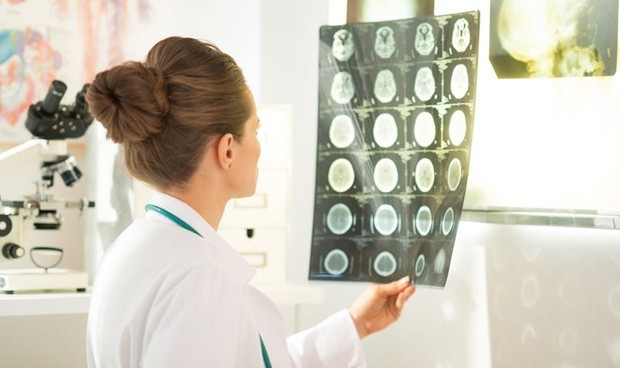
50 to 60 million new cases are being reported in the world. head trauma (TCE) per year, which is estimated to be more than 90 percent are lightand currently computed tomography Cranial CT is the standard diagnostic tool to evaluate intracranial injury in patients with traumatic brain injury to some extent. However, today there are no universally accepted standards When should an urgent head CT be performed in mild TBI. And the use of these protocols at the Spanish level is dependent on the Centre, which means that There is no uniformity among the autonomous communitiesHealth regions and centers of the same region regarding the use of a consensus, pathway, protocol, guide or manual of action for the management of patients with mild TBI.
For this reason, from the Spanish Society of Medical Radiology (serum), with its emergency section (Serau), Spanish Society of Emergency Medicine (Semes), Spanish Society of Laboratory Medicine (seqcml), Spanish Society of Neurosurgery (Cenac) and the Spanish Association of Football Team Doctors have developed, participated in and endorsed a consensus on the management of patients with mild head trauma.
Specifically, in Europe it is estimated that at least two and a half million new cases of TBI occur each year and the age-adjusted incidence of patients with TBI admitted to hospitals is in the middle. 200-300 per 100,000 inhabitants per yearWith wide variations between countries.
Mild TBI is any trauma to a craniocerebral region that raises suspicion of acute brain injury using the WHO diagnostic criteria to identify it. Nowadays, head CT is somewhat of a standard diagnostic tool to evaluate intracranial injury of patients with severe head trauma and identify those who require immediate surgical treatment. Only 7-10 percent of patients present with mild TBI intracranial abnormalities Detected by CT, less than 1 percent of which require surgical intervention and mortality can be classified as Extraordinary (0.1 percent).
There is no consensus on the approach to mild stroke
There is consensus on whether to perform head CT in patients with moderate or severe TBI, but there is no consensus on why patients with mild TBI should undergo this test. Low prevalence of intracranial anomalies Uncommon mortality detected by CT and associated with mild brain damage.
This lack of consensus further increased the need for more objective tools There has been a rapid increase in requests for cranial CT from the emergency department to determine the neurocognitive status of these patients.
For Agustina Vicente And ines pecharomanSERAM’s emergency expert, “Resources need to be optimized more detailed stratification of risk to define the best approach for each patient. Furthermore, with increased associated costs, there has been saturation of services involved and increased risks of radiation exposure (particularly significant for those under 20 years of age), leading to widespread use of immediate head CT in TBI. The usage has been questioned. “And there is a need to adapt.”
Proof of the exponential growth is the recent receipt of CE marking (European Conformity) and approval fda (Food and Drug Administration) The first rapid serum/plasma assay of specific biomarkers GFAP and UCH-L1 in mild TBI.
The results suggest this test could be incorporated into the standard of care to help decision making During evaluation of adult patients with GCS 13-15 in the first 12 hours after injury, to determine the need for CT. “This situation offers the possibility to propose an updated algorithm to standardize the management of mild TBI in emergency situations in Spain,” say Vicente and Pecheroman.
How and when to proceed in patient assessment
When the patient arrives at the emergency room by ambulance or on his own, and life-threatening conditions, multiple trauma, or more severe forms of TBI have been ruled out, for which specific protocols are available, patients with suspected mild TBI are treated. goes less important category, It is evaluated for the purpose of identifying the presence of signs, symptoms, and/or risk factors for intracranial injury. Feeling of Neuroimaging testsThis is limited to patients in whom the risk is high, bearing in mind that in the context of mild TBI, approximately 90 percent of cranial CT scans requested are normal.
Rapid Serum/Plasma Test Specific biomarkers during GFAP and UCH-L1 evaluation in the first 12 hours after trauma are a complementary tool to help in decision making to address the need to perform head CT in patients with symptoms and/or risk factors for GCS 15 . , GCS 14 or GCS 13.
A negative test result is associated with Absence of intracranial lesions Due to its high negative predictive value. Therefore, after negative results in determination of GFAP and UCH-L1, patients can be discharged for home monitoring until the patient recovers and has no symptoms. If more than 12 hours have passed since the trauma or the biomarker result is positive, a blood test is performed. skull whistle, In the event of pathological findings on CT or in a situation when the patient’s symptoms do not agree with the radiological results, the neurosurgery service is consulted to proceed with the evaluation of the patient.
patients with type A CT without pathological findings People who do not have risk factors and who have not experienced clinical deterioration or persistence of symptoms can be discharged to home monitoring. In addition, recommended actions following CT results with and without pathological findings are specified.
What are the causes of suffering a stroke?
The causes of TBI are mostly falls and traffic or work accidents, and to a lesser extent blows to the head, physical violence and contact sports, among other causes.
In clinical practice, it is estimated that approximately 60–70 percent of mild TBI cases involve patients 60 or 65 years of age and older. Previous comorbiditiesof which injury mechanism This is mostly a fall from its height (60–82 percent). The other 30 percent, mainly, correspond to young patients who suffer TBI during physical activity.
Although it may include statements, data, or notes from health institutions or professionals, the information contained in medical writing is edited and prepared by journalists. We advise the reader to consult a health care professional with any health related questions.

